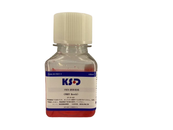
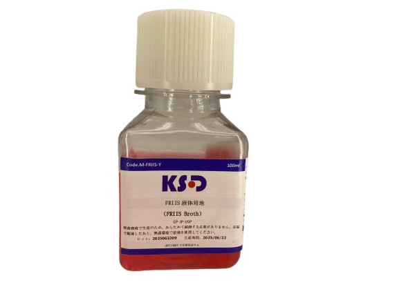
Friis agar is specifically formulated for the cultivation of fastidious avian mycoplasmas and is supplemented with nicotinamide adenine dinucleotide (NAD⁺) to support the metabolic requirements of Mycoplasma synoviae. The medium contains phenol red as a pH indicator, which undergoes a colorimetric shift from red to yellow or purple in response to acidic or alkaline metabolic byproducts, thereby serving as a qualitative marker for mycoplasma growth.
To suppress non-mycoplasmal microbial contaminants, penicillin G is incorporated into the formulation, selectively inhibiting the proliferation of accompanying bacterial flora. Additionally, the supplementation of lipid components from yolk, including cholesterol and essential fatty acids, significantly enhances the growth performance of lipid-requiring strains—most notably Mycoplasma pneumoniae ATCC 23714—by facilitating membrane synthesis and metabolic activity under in vitro conditions.
To prevent growth inhibition caused by the degradation of NAD⁺ during later stages, alternative NAD⁺ analogs are co-supplemented in the culture system.
Microscopic observation is the global gold standard for microbial detection. Although PCR is widely used, a lack of comprehensive mycoplasma research means many commercial kits suffer from false positives and negatives. Thus, agar plate microscopy is still crucial.
At 4x objective and 10x eyepiece magnification, all mycoplasma colonies exhibit a characteristic “fried egg” morphology. At 100x magnification, species-level identification is often possible.
If it's difficult to distinguish mycoplasma from cell debris, use Kusada ready-to-use mycoplasma stain. After staining, rinsing, and resting for 30–60 minutes, debris will fade, while mycoplasma retains a blue color with clearer “fried egg” morphology.
Mycoplasma synoviae is arguably the most difficult strain to culture, with strict dependence on NAD (nicotinamide adenine dinucleotide) for initiating growth pathways. NAD is highly unstable, leading to declining medium efficacy after 3–4 days.
Solutions include periodic NAD supplementation or maintaining NAD stability in both liquid and agar formulations.
Slight yellowing within the first 24 hours is mainly due to changes in ionization from temperature increase, which lowers pH. As incubation continues, sugar degradation further drops the pH, reaching around 7.4, which activates oral mycoplasma growth. Once activated, they produce alkali, shifting the color toward red or reddish-purple.
If the strain is highly active or the inoculum is large, the shift to red may be fast, masking the initial drop in pH.
If the medium turns bright yellow directly, it may indicate weak strain viability or low inoculum size, suggesting SOP optimization is needed—especially if yellowing occurs within a week, which likely indicates a flaw in the liquid medium formulation.
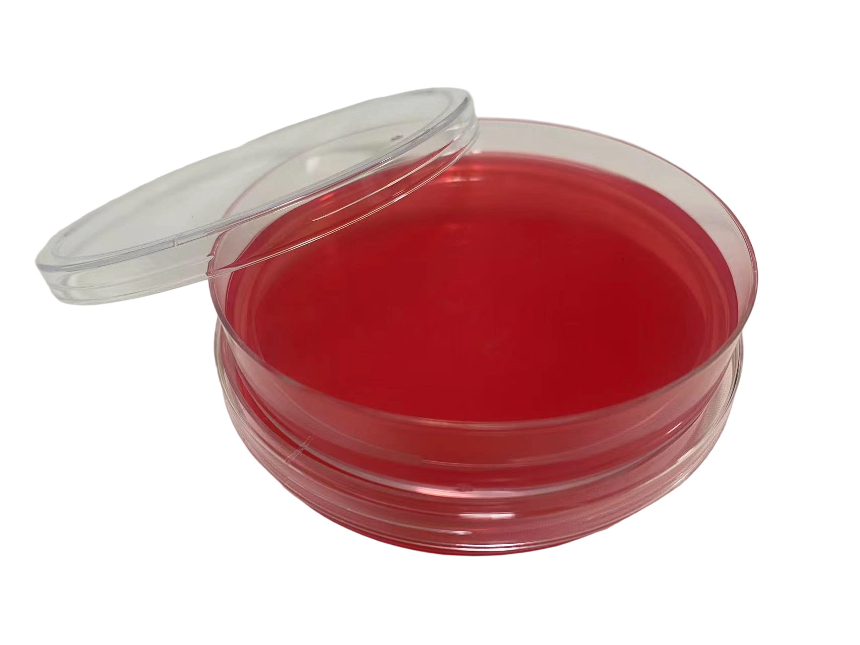
suitable for microbiology, plate diam. 60 mm, For general mycoplasma detection, pack of 10 plates,box of 10 packs
suitable for microbiology, plate diam. 60 mm, For general mycoplasma detection, pack of 10 plates,box of 10 packs
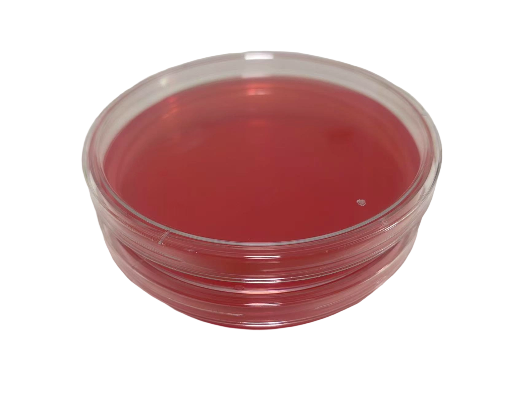
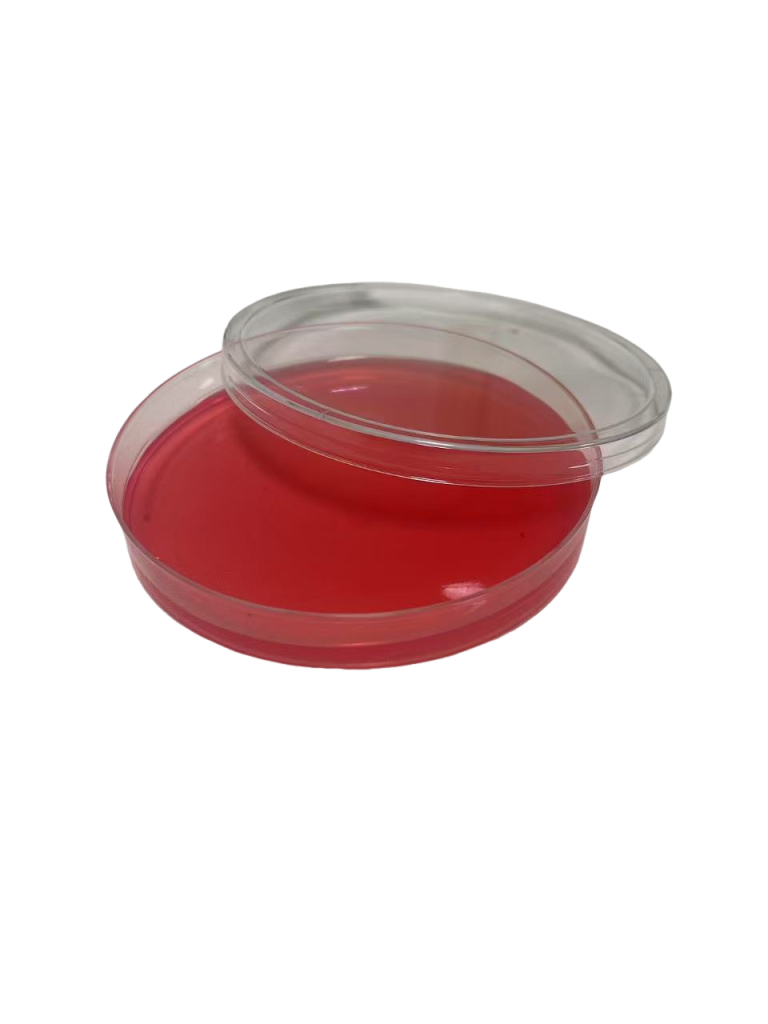
suitable for bioburden testing, For mycoplasma detectionsuitable for bioburden testing, For mycoplasma detectionsuitable for bioburden testing, For mycoplasma detectionsuitable for bioburden testing, For mycoplasma detectionsuitable for bioburden testing, For mycoplasma detectionsuitable for bioburden testing, For mycoplasma detectionsuitable for bioburden testing, For mycoplasma detectionsuitable for bioburden testing, For mycoplasma detection
suitable for bioburden testing, For mycoplasma detection
suitable for bioburden testing, For mycoplasma detection
An additive suitable for cell and mycoplasma culture, at a concentration 10 times higher than the conventional concentration.
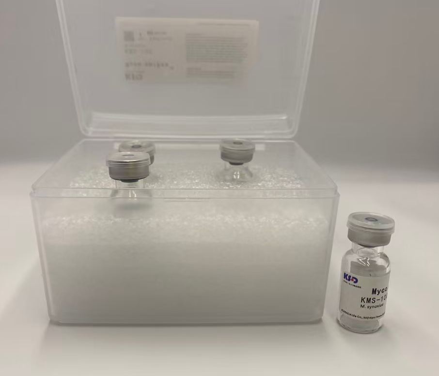
Mycoplasma gallisepticum
Mycoplasma salivarium